
Search
- Page Path
- HOME > Search
Original Article
- Clinical Study
- Relationships between Thigh and Waist Circumference, Hemoglobin Glycation Index, and Carotid Plaque in Patients with Type 2 Diabetes
- Myung Ki Yoon, Jun Goo Kang, Seong Jin Lee, Sung-Hee Ihm, Kap Bum Huh, Chul Sik Kim
- Endocrinol Metab. 2020;35(2):319-328. Published online June 24, 2020
- DOI: https://doi.org/10.3803/EnM.2020.35.2.319
- 8,381 View
- 145 Download
- 4 Web of Science
- 3 Crossref
-
 Abstract
 PDF
PDF PubReader
  ePub
ePub - Background
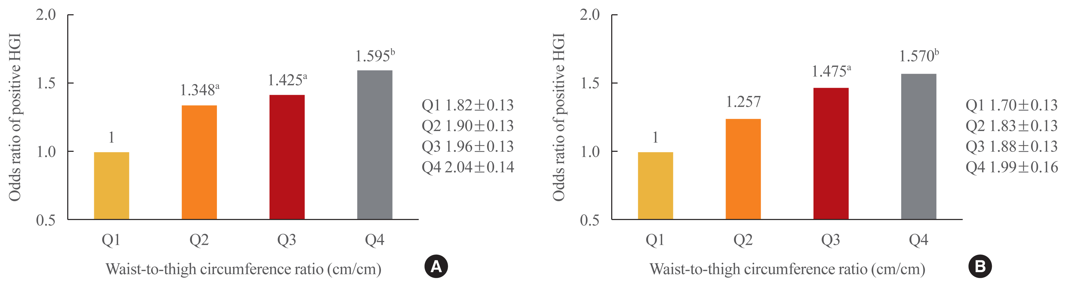
This study investigated the relationships of thigh and waist circumference with the hemoglobin glycation index (HGI) and carotid atherosclerosis in patients with type 2 diabetes.
Methods
This observational study included 3,075 Korean patients with type 2 diabetes, in whom anthropometric measurements and carotid ultrasonography were conducted. HGI was defined as the measured hemoglobin A1c (HbA1c) level minus the predicted HbA1c level, which was calculated using the linear relationship between HbA1c and fasting plasma glucose levels. Carotid atherosclerosis was defined as a clearly isolated focal plaque or focal wall thickening >50% of the surrounding intima-media thickness.
Results
The frequency of a positive HGI decreased with increasing thigh circumference in men and increased with increasing waist circumference in women after adjusting for potential confounding variables. Thigh and waist circumference had a combined augmentative effect on the likelihood of positive HGI, which was dramatically higher in patients in higher waist-to-thigh ratio quartiles (adjusted odds ratios for the highest compared to the lowest quartile: 1.595 in men and 1.570 in women). Additionally, the larger the thigh circumference, the lower the risk of carotid atherosclerosis, although in women, this relationship lacked significance after adjustment for potential confounders.
Conclusion
HGI was associated with thigh circumference in men and waist circumference in women. In addition, the combination of low thigh circumference and high waist circumference was strongly associated with a higher HGI in Korean patients with type 2 diabetes. In particular, thigh circumference was associated with carotid atherosclerosis in men. However, further longitudinal studies are warranted. -
Citations
Citations to this article as recorded by
- Association between hemoglobin glycation index and subclinical myocardial injury in the general population free from cardiovascular disease
Zhenwei Wang, Yihai Liu, Jing Xie, Nai-Feng Liu
Nutrition, Metabolism and Cardiovascular Diseases.2022; 32(2): 469. CrossRef - Association of Hemoglobin Glycation Index With Contrast-Induced Acute Kidney Injury in Patients Undergoing Coronary Angiography: A Retrospective Study
Zhezhe Chen, Duanbin Li, Maoning Lin, Hangpan Jiang, Tian Xu, Yu Shan, Guosheng Fu, Min Wang, Wenbin Zhang
Frontiers in Physiology.2022;[Epub] CrossRef - Associations of continuous glucose monitoring-assessed glucose variability with intima-media thickness and ultrasonic tissue characteristics of the carotid arteries: a cross-sectional analysis in patients with type 2 diabetes
Naohiro Taya, Naoto Katakami, Tomoya Mita, Yosuke Okada, Satomi Wakasugi, Hidenori Yoshii, Toshihiko Shiraiwa, Akihito Otsuka, Yutaka Umayahara, Kayoko Ryomoto, Masahiro Hatazaki, Tetsuyuki Yasuda, Tsunehiko Yamamoto, Masahiko Gosho, Iichiro Shimomura, Hi
Cardiovascular Diabetology.2021;[Epub] CrossRef
- Association between hemoglobin glycation index and subclinical myocardial injury in the general population free from cardiovascular disease

Case Report
- Adrenal gland
- Adult Multisystem Langerhans Cell Histiocytosis Presenting with Central Diabetes Insipidus Successfully Treated with Chemotherapy
- Jung-Eun Choi, Hae Ri Lee, Jung Hun Ohn, Min Kyong Moon, Juri Park, Seong Jin Lee, Moon-Gi Choi, Hyung Joon Yoo, Jung Han Kim, Eun-Gyoung Hong
- Endocrinol Metab. 2014;29(3):394-399. Published online September 25, 2014
- DOI: https://doi.org/10.3803/EnM.2014.29.3.394
- 3,989 View
- 31 Download
- 6 Web of Science
- 5 Crossref
-
Abstract
PDFPubReader
We report the rare case of an adult who was diagnosed with recurrent multisystem Langerhans cell histiocytosis (LCH) involving the pituitary stalk and lung who present with central diabetes insipidus and was successfully treated with systemic steroids and chemotherapy. A 49-year-old man visited our hospital due to symptoms of polydipsia and polyuria that started 1 month prior. Two years prior to presentation, he underwent excision of right 6th and 7th rib lesions for the osteolytic lesion and chest pain, which were later confirmed to be LCH on pathology. After admission, the water deprivation test was done and the result indicated that he had central diabetes insipidus. Sella magnetic resonance imaging showed a mass on the pituitary stalk with loss of normal bright spot at the posterior lobe of the pituitary. Multiple patchy infiltrations were detected in both lung fields by computed tomography (CT). He was diagnosed with recurrent LCH and was subsequently treated with inhaled desmopressin, systemic steroids, vinblastine, and mercaptopurine. The pituitary mass disappeared after two months and both lungs were clear on chest CT after 11 months. Although clinical remission in multisystem LCH in adults is reportedly rare, our case of adult-onset multisystem LCH was treated successfully with systemic chemotherapy using prednisolone, vinblastine, and 6-mercaptopurine, which was well tolerated.
-
Citations
Citations to this article as recorded by- Adult localized Langerhans cell histiocytosis: A case report
Pan-Pan Yang, Su-Ye Hu, Xu-Ya Chai, Xiao-Meng Shi, Li-Xia Liu, Ling-E Li
World Journal of Clinical Cases.2023; 11(34): 8164. CrossRef - Adult Langerhans Cell Histiocytosis Masquerading as Hidradenitis Suppurativa
Jason Chertoff, Julian Chung, Ali Ataya
American Journal of Respiratory and Critical Care Medicine.2017; 195(8): e34. CrossRef - Articles in 'Endocrinology and Metabolism' in 2014
Won-Young Lee
Endocrinology and Metabolism.2015; 30(1): 47. CrossRef - Pulmonary Langerhans Cell Histiocytosis in an Adult Male Presenting with Central Diabetes Insipidus and Diabetes Mellitus: A Case Report
Yeun Seoung Choi, Jung Soo Lim, Woocheol Kwon, Soon-Hee Jung, Il Hwan Park, Myoung Kyu Lee, Won Yeon Lee, Suk Joong Yong, Seok Jeong Lee, Ye-Ryung Jung, Jiwon Choi, Ji Sun Choi, Joon Taek Jeong, Jin Sae Yoo, Sang-Ha Kim
Tuberculosis and Respiratory Diseases.2015; 78(4): 463. CrossRef - Adult-onset Langerhans cell histiocytosis presenting with adipsic diabetes insipidus, diabetes mellitus and hypopituitarism: A case report and review of literature
Erick S. Mendoza, Amy A. Lopez, Valerie Ann U. Valdez, Jean D. Uy-Ho, Sjoberg A. Kho
Journal of Clinical and Translational Endocrinology: Case Reports.2015; 1(1): 1. CrossRef
- Adult localized Langerhans cell histiocytosis: A case report
Original Article
- Impact of Serum Adiponectin Concentration on Progression of Carotid Atherosclerosis in Patients with Type 2 Diabetes Mellitus.
- Chul Sik Kim, Ju Ri Park, Sung Hoon Yu, Jun Goo Kang, Ohk Hyun Ryu, Seong Jin Lee, Eun Gyung Hong, Doo Man Kim, Jae Myung Yoo, Sung Hee Ihm, Moon Gi Choi, Hyung Joon Yoo
- Endocrinol Metab. 2012;27(1):31-38. Published online March 1, 2012
- DOI: https://doi.org/10.3803/EnM.2012.27.1.31
- 2,093 View
- 25 Download
-
Abstract
PDF
- BACKGROUND
Increased cardiovascular events, which is the leading cause of death in type 2 diabetic patients, are mainly caused by accelerated atherosclerosis. Adiponectin has been suggested as a risk factor for cardiovascular diseases in cross-sectional studies. However, little is known about the impact of adiponectin on the progression of carotid atherosclerosis in type 2 diabetic patients. This study was conducted to evaluate the impact of early adiponectin levels on the progression of carotid atherosclerosis. METHODS: From March 2009, 150 patients with type 2 diabetes were consecutively enrolled in our affiliated outpatient clinic. Anthropometric and biochemical data, including adiponectin levels, were measured in each participant. We measured the carotid intima-media thickness (CIMT) at baseline and at 1-year follow-up (n = 111). Then, we prospectively studied the relationship between the serum adiponectin levels and the progression of CIMT for 1 year. RESULTS: Adiponectin levels negatively correlated with CIMT (r = -0.219, P = 0.015). Moreover, mean progression of CIMT was 0.016 +/- 0.040 mm. However, there was no correlation between adiponectin levels and the progression of CIMT within 1-year follow-up period (r = -0.156, P = 0.080). Age (beta = 0.556, P = 0.004), LDL cholesterol (beta = 0.276, P = 0.042), and A1C (beta = 0.309, P = 0.038) were found to be independent risk factors for CIMT. However, A1C (beta = 0.311, P = 0.042) was found to be the only independent risk factor for the progression of CIMT. CONCLUSION: In our study, adiponectin levels were negatively associated with CIMT. However, it did not affect the progression of CIMT at 1-year follow-up. Overall glycemic control is the most important factor in the progression of CIMT in patients with type 2 diabetes.
Case Reports
- A Case of Thyrotoxic Periodic Paralysis with Rhabdomyolysis.
- Seo Hee Lee, Seong Yeol Kim, Hae Ri Lee, Jun Goo Kang, Ohk Hyun Ryu, Chul Sik Kim, Byung Wan Lee, Seong Jin Lee, Eun Gyoung Hong, Hyeon Kyu Kim, Doo Man Kim, Jae Myung Yu, Sung Hee Ihm, Moon Gi Choi, Hyung Joon Yoo
- J Korean Endocr Soc. 2008;23(6):425-429. Published online December 1, 2008
- DOI: https://doi.org/10.3803/jkes.2008.23.6.425
- 1,872 View
- 21 Download
-
Abstract
PDF
- Hyperthyroidism combined with rhabdomyolysis is extremely rare. There are only 6 reported cases of hyperthyroidism accompanied with rhabdomyolysis in the medical literature. Rhabdomyolysis is a syndrome involving the breakdown of skeletal muscle, and this causes myoglobin and intracellular protein to leak into the circulation. The causes of rhabdomyolysis include trauma, electrolyte abnormality, infection, drug, toxin and hypothyroidism. We report here on a patient who presented with thyrotoxic periodic paralysis and rhabdomyolysis with hypokalemia. He complained of lower leg paralysis along with muscle tenderness, and the laboratory findings showed elevated creatine kinase (CK) levels. After treatment by hydration, potassium replacement and drug medication, including propylthiouracil and beta-blocker, his CK levels were normalized and his symptoms were much improved. For patient with thyrotoxic periodic paralysis and muscle tenderness, the possibility of rhabdomyolysis should be clarified by examining the CK levels.
- A Case of Unilateral Exophthalmos Caused by a Dural Arteriovenous Malformation in Thyroid-Associated Ophthalmopathy.
- Sun Ryoung Choi, Seong Jin Lee, Hae Ri Lee, Jun Goo Kang, Ohk Hyun Ryu, Chul Sik Kim, Byung Wan Lee, Eun Gyung Hong, Hyeon Kyu Kim, Doo Man Kim, Jae Myung Yoo, Sung Hee Ihm, Moon Gi Choi, Hyung Joon Yoo
- J Korean Endocr Soc. 2008;23(1):51-55. Published online February 1, 2008
- DOI: https://doi.org/10.3803/jkes.2008.23.1.51
- 1,978 View
- 20 Download
- 2 Crossref
-
Abstract
PDF
- Thyroid-associated ophthalmopathy is associated with thyroid dysfunction, particularly Graves' disease, and is manifested as eye signs, including proptosis. In cases of unilateral exophthalmos with thyroid-associated ophthalmopathy, other causes such as orbital neoplasm, carotid-cavernous fistula, cavernous sinus thrombosis, and dural arteriovenous malformation (AVM) should be excluded. Dural AVM, an abnormal dural arteriovenous connection, is a rare neurovascular entity that mimics thyroid-associated ophthalmopathy. When eye involvement is unilateral or asymmetric, dural AVM can be considered in the differential diagnosis of thyroid-associated ophthalmopathy. A twenty-six year-old woman presented with unilateral exophthalmos in Graves' disease. By orbital magnetic resonance imaging and cerebral angiography, thyroid-associated ophthalmopathy and dural AVM were diagnosed. The unilateral exophthalmos improved after coil embolization of the dural AVM. In summary, we report the first case of a dural AVM with Graves' disease and thyroid-associated ophthalmopathy.
-
Citations
Citations to this article as recorded by- Proptosis as a Primary Symptom of Brain Arteriovenous Malformation
Jong Eun Lee, Jin Sook Yoon, Keun Young Park
Ophthalmic Plastic & Reconstructive Surgery.2020; 36(2): e53. CrossRef - Ophthalmopathy Induced by Bilateral Carotid Cavernous Fistula in a Patient with Graves' Disease
Jong Kun Ha, Ji Hye Suk, A Ra Jo, Chan Woo Jung, Bong Jae Kim, Seong Oh Park, Sang Su Kim, Mi Kyung Kim
Endocrinology and Metabolism.2011; 26(4): 335. CrossRef
- Proptosis as a Primary Symptom of Brain Arteriovenous Malformation
Original Article
- The Clinical Significance of Retinoic Acid Receptor beta Expressions in Primary and Recurred Metastatic Lymph Node Papillary Thyroid Carcinomas.
- Jae Pil Han, Seong Jin Lee, Kyung Chan Choi, Young Euy Park, Hae Ri Lee, Jun Goo Kang, Ohk Hyun Ryu, Chul Sik Kim, Byung Wan Lee, Eun Gyung Hong, Hyeon Kyu Kim, Doo Man Kim, Jae Myung Yoo, Sung Hee Ihm, Hyung Joon Yoo, Moon Gi Choi
- J Korean Endocr Soc. 2007;22(6):419-427. Published online December 1, 2007
- DOI: https://doi.org/10.3803/jkes.2007.22.6.419
- 1,760 View
- 16 Download
-
Abstract
PDF
- BACKGROUND
The present study was designed to investigate the correlations of retinoic acid receptor beta(RARbeta) expression for primary and recurred metastatic lymph node (LN) papillary thyroid carcinoma (PTC) tissues and the correlations of RARbeta expression with the uptake of I(131) as detected on a whole body scan (WBS). METHODS: Primary and metastatic LN PTC tissues were examined by immunohistochemical methods. Staining positivity was calculated, and staining intensity was graded as negative (0), weak (1+), moderate (2+) and strong (3+). Nuclear staining intensity (NSI) of cells from tissues was also examined. RESULTS: Seventeen patients who had regional cervical LN metastasis without distant metastasis were included in the study, and 13 patients had the abnormal uptake of I(131) as detected on a WBS. In primary PTC tissues, RARbeta staining positivity and intensity of carcinoma cells were significantly higher than those of normal cells but NSI was significantly higher in normal cells than carcinoma cells. Between primary and metastatic LN PTC tissues, RARbeta staining intensity was correlated after controlling for age. Primary PTC tissues from 14 (82.4%) out of 17 patients were concordant between NSI and the uptake of I(131) as detected on a WBS. NSI predicted the I(131) uptake as detected on a WBS with 81.3% positive predicted value (PPV) and 100% negative predicted value. Metastatic LN PTC tissues from 13 (76.5%) out of 17 patients were concordant between NSI and the uptake of I(131) as detected on a WBS. NSI predicted the uptake of I(131) as detected on a WBS with 76.5% PPV. When the results of NSI taken either as positive or negative were correlated with those of the uptake of I(131) as detected on a WBS in primary and metastatic LN PTC tissues, the correlation was not significant after controlling for age. CONCLUSION: Our results demonstrate that nuclear RARbeta expression may be decreased in PTC tissues than normal thyroid tissues, and RARbeta expression in primary PTC tissues as well as in recurred metastatic LN PTC tissues may predict the uptake of I(131) as detected on a WBS.
Case Report
- A Novel Mutation of the Vasopressin-Neurophysin II Gene in a Familial Neurohypophyseal Diabetes Insipidus.
- Mi Jung Kim, Byung Wan Lee, In Kyung Jeong, Jun Goo Kang, Seong Jin Lee, Eun Gyung Hong, Hyeon Kyu Kim, Doo Man Kim, Jae Myung Yoo, Sung Hee Ihm, Moon Gi Choi, Hyung Joon Yoo
- J Korean Endocr Soc. 2007;22(2):118-124. Published online April 1, 2007
- DOI: https://doi.org/10.3803/jkes.2007.22.2.118
- 2,044 View
- 21 Download
-
Abstract
PDF
- Autosomal dominant familial neurohypophyseal diabetes insipidus (adFNDI) is a rare form of central diabetes insipidus (DI), and this malady is clinically characterized by polydipsia and polyuria, and it is caused by mutation in the vasopressin-neurophysin II. We identified a Korean family that suffered with adFNDI and we found a novel mutation in the NP II molecule. The index subject's DI symptoms dated to childhood, and his familial history was consistent with autosomal transmission. The diagnosis of central DI was done by performing a water deprivation test and a vasopressin challenge test. For molecular analysis, the genomic DNA was extracted and the AVP-NP II gene was amplified by polymerase chain reaction from four clinically-affected members and seven clinically-nonaffected members. Genetic analysis of AVP-NP II revealed new a heterozygous missense mutation in exon 2 of the AVP-NP II gene (+1692C > A) and this amino acid substitution (Cys105Stop) was predicted to have occurred in four clinically-affected subjects. In summary, in the present study we have described a novel mutation of the AVP-NPII gene in a Korean family suffering with adFNDI.
Original Articles
- The Changes in Atherosclerotic Markers and Adiopocytokines after Treatment with Growth Hormone for the Patients with Hypopituitarism and Growth Hormone Deficiency.
- Hyun Won Shin, In Kyung Jeong, Goo Yeong Cho, Cheul Young Choi, Jong Yeop Kim, Yeong Je Chae, Min Ho Cho, Byung Wan Lee, Seong Jin Lee, Chul Young Park, Eun Gyoung Hong, Hyeon Kyu Kim, Doo Man Kim, Jae Myung Yu, Sung Hee Ihm, Moon Ki Choi, Hyung Joon Yoo, Sung Woo Park
- J Korean Endocr Soc. 2006;21(6):515-525. Published online December 1, 2006
- DOI: https://doi.org/10.3803/jkes.2006.21.6.515
- 1,892 View
- 22 Download
- 1 Crossref
-
Abstract
PDF
- BACKGROUND
It is known that patients with hypopituitarism have a high mortality rate due to the presence of atherosclerosis, cardiovascular diseases and stroke. The aim of this study was the effect of growth hormone (GH) on the atherosclerotic markers and the adipocytokine levels. METHOD: The study was conducted on 13 adult patients with hypopituitarism and growth hormone deficiency (GHD), and they had been stabilized after receiving hormone replacement therapy for other insufficient pituitary hormones, other than GH, for more than one year. Before treatment with GH, we compared the lipid metabolism, glucose metabolism, cardiovascular risk factors and adipocytokine levels, including adiponectin, leptin, TNF-alpha and IL-6, between the GHD patients and 13 healthy adults who were of a similar age and gender distribution. Patients with GHD were treated with 1 U/day of GH for 6 months. We measured insulin-like growth factor-I (IGF-I), blood pressure, body composition, lipid metabolism, glucose metabolism and hs-CRP, cardiac function, adiponectin, leptin, TNF-alpha and IL-6 levels, flow mediated vasodilation (FMD) and nitroglycerin mediated vasodilation (NMD) before and after GH treatment. RESULTS: The patients with hypopituitarism and GHD showed significantly higher levels of total cholesterol (P = 0.002), low-density lipoprotein cholesterol (LDL-C) (P = 0.036), hs-CRP (P = 0.0087) and leptin (P < 0.001) than did the normal healthy adults. However, there was no difference between the normal adults and the patients with GHD for the systolic and diastolic BP, the levels of apoA, apoB, fasting blood glucose(FBG) and HOMA-IR. In the subjects with GHD after treatment with GH, the level of fat mass (P = 0.0017), total cholesterol (P = 0.004), LDL-C (P = 0.001), leptin (P = 0.013), TNF-alpha (P < 0.001) and hs-CRP (P = 0.0001) were significantly reduced, while lean body mass (P = 0.0161), FFA (P = 0.049) and FMD (P = 0.0051) showed a significant increase. However, there was no significant difference in the level of the systolic and diastolic BP, LDL-C, apoA, apoB, LP (a), HOMA-IR, ejection fraction, left ventricular posterior wall, E/A ratio, intraventricular septum, NMD, intima-media thickness, adiponectin, IL-6, FBG and fasting insulin before and after GH treatment. CONCLUSION: The subjects with GHD were vulnerable to cardiovascular disease. GH therapy for 6 months had a positive effect on their various cardiovascular risk factors. -
Citations
Citations to this article as recorded by- Molecular Biology of Atherosclerosis
In-Kyung Jeong
Endocrinology and Metabolism.2010; 25(3): 166. CrossRef
- Molecular Biology of Atherosclerosis
- Duration of Preparation for Postoperative Radioiodine Administration in Differentiated Thyroid Carcinoma.
- Hyeon Kyu Kim, Min Ho Cho, Choel Young Park, Seong Jin Lee, Gi Weon Oh, In Kyung Jeong, Eun Gyung Hong, Sung Hee Ihm, Doo Man Kim, Jae Myung Yu, Moon Gi Choi, Hyung Joon Yoo, Sung Woo Park, Jin Hwan Kim, Young Soo Rho
- J Korean Endocr Soc. 2005;20(5):460-466. Published online October 1, 2005
- DOI: https://doi.org/10.3803/jkes.2005.20.5.460
- 1,870 View
- 24 Download
-
Abstract
PDF
- BACKGROUND
Radioiodine treatment is effective for the removal of remnant thyroid tissues after thyroidectomy in patients with differentiated thyroid carcinoma. To induce the elevation of serum TSH level which facilitates the uptake of radioiodine into remnants, a 4 to 6 week interval between thyroidectomy and radioiodine administration has been established. During the period of preparation, most patients have experienced overt symptoms of hypothyroidism which have led to the development of alternative strategies. Some reports have suggested that the interval could be reduced to about 3 weeks with less symptoms. We reevaluated the adequate time needed for the elevation of serum TSH level above 30microU/mL after thyroidectomy. METHODS: Forty five patients who had undergone total thyroidectomy for differentiated thyroid carcinoma were investigated. Serum TSH and free T4 levels were measured one or more times within 3 weeks after operation(total 97 blood samples). Eighty nine blood samples were obtained within 15 days. RESULTS: In 41 patients (91.1%) serum TSH levels increased to 30 microU/mL until 15 days after operation. Until postoperative 21 days, serum TSH levels in all the other patients reached 30microU/mL. In linear equation, the daily increment of serum TSH levels was 2.62microU/mL for the first 8 days after operation and 5.34micorU/mL for the next 7 days. The half-life of serum free T4 levels showed marked individual variations. CONCLUSION: Measurement of serum TSH level at about 15 days after total thyroidectomy for differentiated thyroid carcinoma may be useful in determining the time of radioiodine administration.
Case Reports
- A Case of Hashimoto's Thyroiditis with Transient T3-Thyrotoxicosis Induced by Hydatidiform Mole.
- Ji Youn Yoo, Hong Ju Moon, Cheol Young Park, Seong Jin Lee, In Kyung Jeong, Eun Gyung Hong, Gi Weon Oh, Hyeon Kyu Kim, Doo Men Kim, Jae Myung Yoo, Sung Hee Ihm, Moon Gi Choi, Hyung Joon Yoo, Sung Woo Park, Soo Kee Min
- J Korean Endocr Soc. 2005;20(3):294-298. Published online June 1, 2005
- DOI: https://doi.org/10.3803/jkes.2005.20.3.294
- 1,923 View
- 19 Download
- 1 Crossref
-
Abstract
PDF
- Human chorionic gonadotropin(HCG) is a member of the glycoproteins family synthesized by the placenta, which consists of 2 noncovalently joined subunits(alpha(alpha) and beta(beta)). The alpha- and beta-subunits have a structural homology with the alpha- and beta-subunits of TSH and LH. The thyrotropic action of HCG results from its structural similarity to TSH, so beta-HCG can bind to the TSH receptor in the thyroid gland. A high level of HCG accompanied by an increased thyroid hormone level, can be observed in gestational trophoblastic disease (GTD), such as a hydatidiform mole or a choriocarcinoma, but the clinical symptoms of hyperthyroidism are rarely observed. We experienced a case of Hashimoto's thyroiditis, where the patient was diagnosed with T3-thyrotoxicosis, which had initially been induced by excess beta-HCG due to an H-mole; after evacuation of the H-mole, the condition was diagnosed as hypothyroidism. It has been speculated that a patient with Hashimoto's thyroiditis could have hyperthyroidism, induced by beta-HCG, due to an H-mole
-
Citations
Citations to this article as recorded by- Transient T3 toxicosis associated with Hashimoto’s disease
Sarah Jaroudi, Meredith Gavin, Kathryn Boylan, Alan N. Peiris
Baylor University Medical Center Proceedings.2019; 32(1): 80. CrossRef
- Transient T3 toxicosis associated with Hashimoto’s disease
- A Case of Apical Hypertrohic Cardiomyopathy Associated with Pheochromocytoma.
- Joon Ho Moon, Sung Woo Park, Sung Hee Ihm, Cheol Young Park, Ki Won Oh, Cheol Soo Choi, Seong Jin Lee, In Kyung Jung, Eun Gyung Hong, Hyeon Kyu Kim, Doo Man Kim, Jae Myung Yoo, Moon Gi Choi, Hyung Joon Yoo, So Young Ku, Soo Kee Min
- J Korean Endocr Soc. 2004;19(5):522-527. Published online October 1, 2004
- 1,134 View
- 18 Download
-
Abstract
PDF
- Pheochromocytomas often present with cardiovascular manifestations, such as arrhythmia, angina pectoris and acute myocardial infarction and so on. Both dilated and nonobstructive hypertrophic cardiomyopathies are also rare complications of pheochromocytomas. In hypertrophic cardiomyopathy, an apical variant form constitutes about 25% of cases in Japan, but only 1 to 2% of those in non-Japanese populations, including Korea. The cause of apical hypertrophic cardiomyopathy (AHC) remains unknown. Recently, some cases of AHC associated with pheochromocytomas have been reported, with catecholamine thought to be an important cause. AHC associated with a pheochromocytoma has never been previously reported in Korea. Herein is reported our experience of a case of apical hypertrophic cardiomyopathy associated a pheochromocytoma with a review of the literature
- A Case of Masked Hypoglycemia during Lactic Acidosis.
- Hee Seon Kim, Ho Sung Yoon, Chang Ok Koh, Hyeon Kyu Kim, Choel Young Park, Seong Jin Lee, Gi Weon Oh, In Kyung Jeong, Eun Gyung Hong, Cheol Soo Choi, Doo Man Kim, Sung Hee Ihm, Jae Myung Yu, Moon Gi Choi, Hyung Joon Yoo, Sung Woo Park, Dong Jin Oh
- J Korean Endocr Soc. 2004;19(4):406-410. Published online August 1, 2004
- 1,077 View
- 17 Download
-
Abstract
PDF
- Severe hypoglycemia induces neuroglycopenic symptoms, including mental alteration, as glucose is the exclusive fuel for the central nervous system. However, some reports have shown that non-glucose fuels, like lactates and ketones, could be utilized by the brain during severe hypoglycemia. Herein, a case of extreme hypoglycemia in a 44-year old woman, subsequently diagnosed as congestive heart failure accompanied by ischemic hepatitis and lactic acidosis, is presented. In two episodes of extreme hypoglycemia, she was fully alert without obvious neurological deficits. In this unusual case, an increased supply of lactate might have maintained the cerebral function and prevented cerebral injury during the hypoglycemia that was induced as a result of starvation and hepatic and cardiac dysfunctions
- A Case of Adrenocortical Oncocytoma.
- Seong Jin Lee, Ho Gwon Lee, Cheol young Park, In Kyung Jeong, Eun Gyung Hong, Gi Weon Oh, Hyeon Kyu Kim, Doo Man Kim, Jae Myung Yoo, Sung Hee Ihm, Moon Gi Choi, Hyung Joon Yoo, Sung Woo Park
- J Korean Endocr Soc. 2004;19(1):82-89. Published online February 1, 2004
- 1,052 View
- 17 Download
-
Abstract
PDF
- Oncocytomas are neoplasms, histologically are composed of epithelial cells, with abundant, acidophilic and granular cytoplasm. Electron microscopic studies of oncocytomas have shown that the cytoplasm of oncocytes is packed with mitochondria. The adrenal gland is a very rare anatomical site for oncocytomas, and to the best of our knowledge, only thirty-six cases of adrenal oncocytomas have been described. Herein, a case of a large adrenal mass in a forty-year-old man, which was incidentally detected by abdominal ultrasonography, is presented. This patient demonstrated no clinical manifestation associated with adrenal hyperfunction. Hormonal studies showed no abnormal findings, except for a mild elevation of the 24-hour urinary VMA level. Abdominal computed tomography with enhancement revealed a large, well-defined left adrenal mass, measuring 5.0x.2 x.0cm. The patient underwent a left adrenalectomy, and a light microscopic examination confirmed an adrenocortical oncocytoma, with characteristic oncocytes and polygonal, abundant, eosinophilic and granular cytoplasm. The tumor cells were positive for cytokeratin and vimentin as well as S-100, but negative for chromogranin on immunohistochemical staining. An electron microscopic examination demonstrated closely packed mitochondria, containing intramitochondrial inclusions. After surgery, there was no evidence of a recurrent or distant metastatic disease at the 5 month follow-up. In summary, an extremely rare case of a man with an adrenocortical oncocytoma is reported, which was confirmed by histological examinations, including electron microscopy.
Original Articles
- The Change of Pulmonary Artery Pressure in Graves'Disease Before and After Treatment.
- Taek Man Nam, Han Soo Cho, Jin Seo Lee, Young Rim Song, Doo Man Kim, Young Cheoul Doo, Cheol Young Park, In Kyung Jeong, Eun Gyung Hong, Seong Jin Lee, Gi Weon Oh, Hyeon Kyu Kim, Jae Myung Yu, Moon Gi Choi, Hyung Joon Yoo, Sung Woo Park
- J Korean Endocr Soc. 2003;18(5):465-472. Published online October 1, 2003
- 1,126 View
- 19 Download
-
Abstract
PDF
- BACKGROUND
Exertional symptoms, dyspnea and impaired effort tolerance are common in patients with Graves' disease. Proposed explanations include: high-output left heart failure, ineffective oxygen utilization and respiratory muscle weakness. In addition, pulmonary hypertension has also been reported in patients with Graves' disease. A high prevalence of hypothyroidism and positive thyroid autoantibody were also observed in patients with pulmonary arterial hypertension. Therefore, the pulmonary artery pressure in patients with Graves' disease was evaluated. METHODS: Two-dimensional and Doppler echocardiographic examinations (Hewlett Packard Sonos 2500) were performed to determine the pulmonary artery (PA) pressure in 26 Graves' disease patients, both before and after treatment (23 patients with propylthiouracil and 3 with RAI), and in 10 euthyroid controls. The changes in the PA pressure after treatment were evaluated in 13 patients with Graves' disease, who became euthyroid after treatment. RESULTS: The pulmonary artery pressure was increased in the untreated Graves' disease patients compared to the normal controls (23.5+/-2.32 vs. 29.6+/-10.3 mmHg). 38.5% of the Graves' disease patients (10/26) showed pulmonary arterial hypertension (PA>30 mmHg) and the serum TBII levelwas higher in the Graves' disease patients with pulmonary arterial hypertension than in those with normal PA pressure (P<0.05). In the Graves' patients who became euthyroid after treatment, the PA pressure was significantly decreased. CONCLUSION: 38.5% of the untreated Graves' disease patients showed pulmonary arterial hypertension, and the pulmonary artery pressure was significantly decreased in those who became euthyroid after treatment. The pathogenesis and clinical importance of pulmonary arterial hypertension in Graves' disease requires further studies.
- The Effect of T3 on Thyroid Hormone Receptor Dynamics in Thyroid Hormone Response Element of Chicken Lysozyme Silencer.
- Seong Jin Lee, Cheol Young Park, In Kyung Jeong, Eun Gyung Hong, Cheol Soo Choi, Hyeon Kyu Kim, Doo Man Kim, Jae Myung Yoo, Sung Hee Ihm, Moon Gi Choi, Hyung Joon Yoo, Sung Woo Park, P Reed Larsen
- J Korean Endocr Soc. 2003;18(4):379-391. Published online August 1, 2003
- 1,101 View
- 17 Download
-
Abstract
PDF
- BACKGROUND
The regulation of gene transcription can be controlled by both positive (enhancer) and negative (silencer) regulatory sequences. Several enhancer and silencer elements have been described in the 5' region of the chicken lysozyme gene. The silencer located at -2.4 kb upstream of the chicken lysozyme gene is composed of two separate modules (F1 and F2) that can function as silencers by themselves, but also show synergistic repression after multimerization. The F1 module is bound by a protein termed NeP1 and F2 module, a F2 thyroid hormone response element (F2-TRE), and can be bound by the thyroid hormone receptor (TR). F2-TRE has an inverted palindromic structure, with high affinity to TR. Although many current reported results have tried to explain the regulatory mechanism of chicken lysozyme gene expression due to the thyroid hormone, there have been few studies that clarify the TR dynamics in the F2-TRE of the chicken lysozyme gene, either with or without exposure of the thyroid hormone. Here, the changes in the TR binding patterns in the F2-TRE of the chicken lysozyme gene are described, both before and after T3 stimulation over time. METHODS: Using the stably transfected rat pituitary somatotroph tumor cell line, GC8 cells, with the F2-TRE inserted 5' to the thymidine kinase (TK) promoter, together with a mouse TRalpha- expressing plasmid, a chromatin immunoprecipitation (ChIP) technique was employed to reveal the TR-TRE interaction before and after T3 stimulation. Following the cross-linking and sonication of the cells, the immunoprecipitation was performed overnight, at 4 degrees C, with TRalpha1, TRbeta1 and TRbeta2 antibodies, respectively. The binding patterns and amounts of TRalpha1, TRbeta1 and TRbeta2 to the F2-TRE, before and after 12 hours of 100 nM T3 stimulation, were analyzed using conventional and quantitative real-time polymerase chain reactions (RQ-PCR). The ChIP technique was used to give a basal value for 20 minutes and 1, 2, 4, 6, 8 and 12 hours after the 100 nM T3 stimulation, and RQ-PCR was then performed. Western blot with TRalpha1, TRbeta1 and TRbeta2 antibodies were also performed. RESULTS: After 12 hours of 100 nM T3 stimulation of the GC8 cells, the TRalpha1 and TRbeta2 binding to the F2-TRE increased, but the TR 1 binding to the F2-TRE decreased, by conventional PCR. Although all the TR isoforms were bound to the F2-TRE by RQ-PCR, the TR 1 binding to the F2-TRE, after 12 hours of 100 nM T3 stimulation, was significantly increased (1.01-->2.73, delta=+170.3%, p<0.05), but the change in the amount of TRbeta2 binding was not significant (2.53-->2.98, delta=+17.8%). The TRbeta1 binding was significantly decreased compared with that of the basal level (4.59-->2.06, delta=-55.1%, p<0.05). The total TR bindings to the F2-TRE had a tendency to decrease after 12 hours of 100 nM T3 stimulation (8.13-->7.77, delta=-4.4%). The binding patterns and amounts of TRalpha1, TRbeta1 and TRbeta2, both before and after the 100 nM T3 stimulation, were also identified over time. While the TRbeta1 bindings to the F2-TRE after 1 hour of 100 nM T3 stimulation were acutely reduced, those of the TRalpha1 at 20 minutes and 6 hours were increased. The TRbeta2 bindings showed a maximal increase at 20 minutes. The directions of the TR binding patterns, between the before and after 2 hours of 100 nM T3 stimulation, were identical to those for between 4 and 6 hours of T3 stimulation. There was no significant difference in the TR bindings to the F2-TRE in relation to the amounts (1.5 vs. 4.5 microliter) of TR antibodies used during the ChIP assays. The Western blots showed no significant change of the levels of each TR isoform proteins, either before or after 12 hours of exposure to 100 nM T3. CONCLUSION: These results show the dynamic binding patterns of the TR isoforms to the F2-TRE of the chicken lysozyme gene, both before and after T3 stimulation, over time. Further investigation, however, will be needed to clarify the mechanisms of our observations. The ChIP technique may then be used to reveal the dynamic models of the cofactors, as well as TR isoforms, in the TR-regulated transcription machinery.
 First
First Prev
Prev


